
Free Shipping on Orders Over $200
The Purest Peptides. Period.
Lab Tested and Shipped from the USA
Posted on

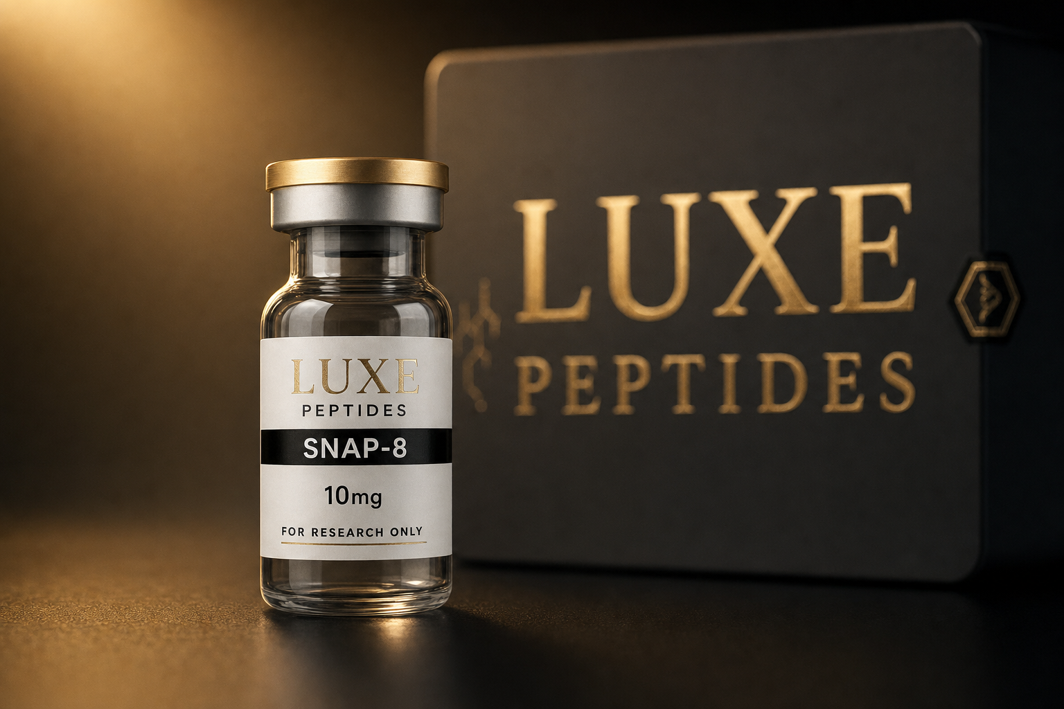
SNAP-8 (Acetyl Octapeptide-3): SNARE Mechanism

Research use only. The material below describes laboratory and preclinical findings for research reference; it is not medical, cosmetic, or human-use guidance, and the compound discussed is not for human or veterinary use.

SNAP-8, catalogued under the INCI designation acetyl octapeptide-3, is an eight-residue synthetic peptide that appears in the ingredient-science and neurobiology literature as a sequence mimetic of the N-terminal region of SNAP-25 — one of the three proteins that assemble into the neuronal SNARE complex. It is one of the more mechanistically specific claims made for a small synthetic peptide, and also one of the more frequently over-stated. This guide covers what the SNARE complex is, what the proposed in vitro mechanism for acetyl octapeptide-3 actually asserts, how that assertion is commonly contrasted with the enzymatic mechanism of botulinum neurotoxin type A (BoNT/A), and where the evidence base is thin. Product reference: SNAP-8 (acetyl octapeptide-3).

The SNARE complex in brief

SNARE proteins (soluble N-ethylmaleimide-sensitive factor attachment protein receptors) are the core machinery of regulated vesicle fusion. In the presynaptic terminal, three partners contribute four helical SNARE motifs to a single parallel four-helix bundle: syntaxin-1A and SNAP-25 on the plasma membrane, and VAMP-2 (synaptobrevin) on the vesicle membrane. SNAP-25 is unusual in contributing two motifs to the bundle rather than one, and it is anchored by palmitoylated cysteines rather than a transmembrane domain.

Assembly is generally modelled as progressive N-to-C “zippering”: the four motifs nucleate at their membrane-distal N-terminal ends and zipper toward the membranes, and the free energy released as the bundle forms is what pulls the two bilayers into contact. Crystallographic work on the assembled ternary complex resolved the bundle’s layered hydrophobic core and the central ionic layer, and biochemical reconstitution established that these three proteins are sufficient to drive fusion in defined systems. The relevant point for any small-molecule or peptide modulator is that the N-terminal nucleation step is the rate-limiting and most interference-susceptible stage of assembly.

Chemistry of acetyl octapeptide-3

The molecule is a linear octapeptide, Ac-Glu-Glu-Met-Gln-Arg-Arg-Ala-Asp-NH2 (single-letter: EEMQRRAD), with an acetylated N-terminus and an amidated C-terminus, giving a molecular weight of approximately 1,075 Da. Both terminal caps neutralise the charges that a free peptide terminus would carry and reduce susceptibility to exopeptidase trimming — a standard stabilisation strategy for short synthetic peptides.

The sequence corresponds to a segment within the N-terminal SNARE motif region of SNAP-25. It is also a two-residue extension of the better-known hexapeptide analogue acetyl hexapeptide-8 (EEMQRR), which shares the same core motif; the added Ala-Asp is the structural difference between the two, and comparative claims between them rest largely on in vitro assay data rather than on independent structural work.

The proposed mechanism: competition, not cleavage

The mechanism attributed to acetyl octapeptide-3 in the ingredient literature is competitive interference with SNAP-25 incorporation into the SNARE complex. The proposal runs as follows: because the peptide reproduces part of the SNAP-25 N-terminal interaction surface, a soluble excess of it is described as engaging the nascent syntaxin/VAMP interface and reducing the efficiency with which full-length SNAP-25 nucleates into the four-helix bundle. Assemblies formed in the presence of the peptide are described as less stable or slower to form, and in vitro assays — typically catecholamine release from chromaffin cells, the classical model system for SNARE-dependent exocytosis — have been reported to show reduced stimulated release under peptide exposure.

Several features of this proposal deserve to be stated plainly, because they are frequently lost in summary:

  • It is stoichiometric, not catalytic. A competitive mimetic must be present in molar excess and occupies its target only while bound. One peptide molecule interferes with, at most, one assembly event.
  • Nothing is cleaved. SNAP-25 is not modified, degraded, or permanently inactivated in this model; the proposed effect is reversible on washout.
  • The buried surface is small. A SNARE motif is on the order of 60–70 residues. An eight-residue fragment reproduces a small fraction of that interface, and the affinity of such a fragment for a partially assembled complex is correspondingly modest.
  • Barrier penetration is a recognised limitation. A ~1,075 Da peptide carrying two arginines and three acidic residues is a poor candidate for passive diffusion across biological membranes, which is why in vitro assay results and formulated-system behaviour are not interchangeable.

Research in this area therefore supports a mechanistic hypothesis with in vitro support, not an established quantitative effect.

Mechanistic contrast with botulinum neurotoxin type A

The comparison to BoNT/A is made constantly, and it is worth being precise about what the two mechanisms share: only the target protein. BoNT/A is a ~150 kDa bacterial protein whose light chain is a zinc-dependent endopeptidase. After receptor-mediated uptake and translocation into the cytosol of a neuron, it cleaves SNAP-25 between Gln197 and Arg198, removing nine C-terminal residues and producing a truncated protein that can no longer support productive SNARE zippering. That is a catalytic, covalent, and functionally durable event; a single light-chain molecule can process many substrate molecules.

Property Acetyl octapeptide-3 (SNAP-8) Botulinum neurotoxin type A (BoNT/A)
Molecular class Synthetic octapeptide, ~1,075 Da Bacterial protein toxin, ~150 kDa
Molecular target N-terminal SNARE assembly interface SNAP-25 polypeptide chain
Mode of action (as described) Competitive occupancy / mimicry Zinc-dependent proteolysis
SNAP-25 covalently altered? No Yes — cleaved at Gln197–Arg198
Stoichiometry Stoichiometric; molar excess required Catalytic; one enzyme, many substrates
Reversibility in vitro Reversible on washout Not reversible; requires protein turnover
Cell entry Passive; poor for a charged peptide Receptor-mediated, high-efficiency
Evidence base Limited in vitro data, largely unreplicated Extensive indexed primary literature

The two mechanisms are placed side by side because they converge on the same protein, not because they are alternatives to one another. No equivalence of potency, kinetics, or biological consequence is implied by the comparison, and none is supported by the available data.

What the evidence base supports — and what it does not

The SNARE biology underlying the hypothesis is exceptionally well established: the complex’s composition, structure, assembly order, and its cleavage by clostridial neurotoxins are all supported by decades of indexed primary literature. The peptide-specific layer is not. Much of the primary in vitro characterisation of acetyl octapeptide-3 and its hexapeptide analogue originates in ingredient-manufacturer technical documentation and conference material rather than independently replicated, peer-reviewed studies, and independent structural evidence of the peptide bound to a partially assembled SNARE complex does not appear to have been published. Researchers designing work in this area should treat the competitive-mimetic model as a testable hypothesis and build in the controls — scrambled-sequence peptide, concentration-response range, washout arm — that the existing datasets largely lack. Analytical documentation for material on hand is in the COA library.

Laboratory handling and arithmetic

Acetyl octapeptide-3 is supplied lyophilised and is readily water-soluble owing to its two glutamates, aspartate, and two arginines. Standard practice for short synthetic peptides applies: store the lyophilised powder cold and desiccated, protect from repeated freeze–thaw cycles once in solution, and account for the methionine residue, which is oxidation-prone and can complicate mass-spectrometric identity checks on aged stock. Concentration arithmetic for stock preparation — mass of peptide over volume of diluent, converted to the molarity an assay protocol calls for — can be worked through with the reconstitution calculator. This is laboratory arithmetic for in vitro work, not administration guidance.

A different mechanistic class for comparison

Researchers surveying this category often place acetyl octapeptide-3 alongside GHK-Cu, but the two share nothing mechanistically. GHK-Cu is a copper-binding tripeptide studied for copper coordination and transcriptional modulation across broad gene sets in cultured cells — a signalling and metal-transport mechanism operating on gene expression timescales, not an interference mechanism acting on a presynaptic protein complex within milliseconds. They belong in the same catalogue, not in the same mechanistic comparison.

References

  • Söllner T, Whiteheart SW, Brunner M, et al. Nature. 1993;362(6418):318–324. PMID: 8455717.
  • Blasi J, Chapman ER, Link E, et al. Nature. 1993;365(6442):160–163. PMID: 8103915.
  • Sutton RB, Fasshauer D, Jahn R, Brunger AT. Nature. 1998;395(6700):347–353. PMID: 9759724.
  • Schiavo G, Matteoli M, Montecucco C. Physiological Reviews. 2000;80(2):717–766. PMID: 10747206.
  • Jahn R, Scheller RH. Nature Reviews Molecular Cell Biology. 2006;7(9):631–643. PMID: 16912714.
  • Südhof TC, Rothman JE. Science. 2009;323(5913):474–477. PMID: 19164740.
  • Blanes-Mira C, Clemente J, Jodas G, et al. International Journal of Cosmetic Science. 2002;24(5):303–310. (Title omitted; the hexapeptide-analogue in vitro study most often cited in this area.)
  • Primary in vitro data specific to acetyl octapeptide-3 appears largely in ingredient-manufacturer technical literature rather than indexed primary research; those datasets are cited generically here because independent replication is not available.

Reviewed for research accuracy — 1 August 2026.

Research use only. This compound is intended solely for in vitro laboratory research and is not for human or veterinary use, diagnostic use, or use in any food, drug, or cosmetic product.
Posted on

Ovagen (Glu-Asp-Leu): A Hepatic Peptide Bioregulator

All compounds discussed here are supplied strictly for in vitro and laboratory research use only; they are not drugs, dietary supplements, or products intended for human or veterinary administration.

Ovagen is one of the most consistently misread entries in the Khavinson short-peptide catalog, and the reason is purely linguistic. The prefix reads as a reference to ovarian tissue, and a large share of the search traffic that reaches pages like this one arrives on that assumption. The assumption is incorrect. In the originating literature, Ovagen is a tripeptide — Glu-Asp-Leu, conventionally written EDL — carrying a hepatic and gastrointestinal tissue designation. Nothing in the source research assigns it to reproductive tissue of any kind.

That mismatch is worth correcting at the top rather than in a footnote. In a family whose members are distinguished from one another only by a three- or four-letter sequence, a name-driven misassumption is the quickest route to characterizing the wrong molecule.

Structure and physical identity

  • Sequence: Glu-Asp-Leu (EDL), unmodified L-amino acids with free N- and C-termini
  • Length: three residues
  • Molecular formula: C15H25N3O8
  • Nominal molecular weight: approximately 375.4 Da for the free peptide; acetate-salt material assays higher, which is why certificate mass and calculated mass rarely agree exactly
  • Charge character: two acidic residues followed by a hydrophobic C-terminal leucine, giving a net-anionic peptide — structurally the opposite pole from the lysine-initiated bioregulators such as Vilon (KE) or Livagen (KEDA)

At roughly 375 Da, EDL is on the order of a tenth the mass of the mid-size research peptides that dominate the rest of the catalog. That size is central to the category’s mechanistic model rather than incidental to it: what the source literature proposes is not sustained receptor occupancy but transient access to the nuclear compartment.

The designation is hepatic and gastrointestinal

Each synthetic peptide in the Khavinson series carries an organ-system assignment inherited from the tissue fraction whose activity it was designed to reproduce, and those assignments are tabulated consistently across the group’s reviews and monographs. EDL appears in those tables under liver and gastrointestinal tract. Reports from this school describe the peptide in hepatocyte and rodent hepatic-tissue models and in gastrointestinal mucosal preparations; the endpoints reported are expression-level and histological rather than clinical, and they have not been reproduced in controlled human research.

Why the name points the wrong way

The names in this catalog are trade designations coined by the developing institute rather than systematic nomenclature, and they are under no obligation to encode the tissue assignment. Some do — Bronchogen and Cardiogen are transparent about it. Ovagen does not, and its resemblance to ovum is unrelated to how the compound is actually classified in the source work. The sequence, not the trade name, is the identifying fact.

The single-residue trap is worth spelling out, because it is the practical failure mode in this family. Ala-Glu-Asp-Leu (AEDL) is Bronchogen, designated for respiratory epithelium. Glu-Asp-Leu (EDL) is Ovagen, designated for hepatic and gastrointestinal tissue. The two share an identical C-terminal tripeptide, differ by a single N-terminal alanine, and are assigned to entirely different organ systems. Nothing about their appearance, solubility, or lyophilized mass distinguishes them at the bench.

Ovagen and Livagen

Livagen is the compound searchers most often pair with Ovagen, and the pairing is reasonable: it is the other liver-designated member of the series. Livagen is Lys-Glu-Asp-Ala (KEDA), a tetrapeptide built on the same basic N-terminal motif as Vilon (KE) and Vesugen (KED) rather than on EDL’s acidic opening. The two are liver-associated by designation but structurally unrelated, and they are not interchangeable in an experimental design.

Livagen also carries the larger primary literature of the two, and most of it concerns chromatin state rather than hepatic tissue directly: cultured human lymphocyte reports describing decondensation of heterochromatin and activation of ribosomal gene clusters following exposure to KEDA. Those are cell-culture endpoints in a small number of reports from a single group, and they are routinely overstated in secondary summaries.

Pancragen (Lys-Glu-Asp-Trp, KEDW) is included in the comparison below as the third member of the digestive-system group these two are usually cross-shopped against; its designation in the same tables is pancreatic tissue.

Comparison

Ovagen Livagen Pancragen
Sequence Glu-Asp-Leu (EDL) Lys-Glu-Asp-Ala (KEDA) Lys-Glu-Asp-Trp (KEDW)
Residues 3 4 4
Formula C15H25N3O8 C18H31N5O9 C26H36N6O9
Nominal MW (free peptide) ~375.4 Da ~461.5 Da ~576.6 Da
Tissue designation in the source literature Liver and gastrointestinal tract Liver Pancreas
Structural class Acidic N-terminus, hydrophobic C-terminus; net anionic Lys-initiated KED- series tetrapeptide Lys-initiated KED- series tetrapeptide, aromatic C-terminus
Where the primary reports concentrate Hepatic and gastrointestinal tissue models Lymphocyte chromatin and gene-activation studies Pancreatic cell and carbohydrate-metabolism models

The proposed mechanism, and its limits

The bioregulator literature assigns these peptides no cell-surface receptor. The proposed site of action is intracellular: fluorescently labeled short peptides have been reported to appear in the nuclei and nucleoli of cultured cells, and the same line of work described sequence-selective binding between labeled short peptides and synthetic deoxyribooligonucleotides in vitro. From those observations the group proposes that a given sequence interacts preferentially with particular promoter-region double-stranded DNA, with molecular-modeling reports suggesting accommodation in the major groove and a sensitivity to CG content and cytosine methylation state.

Applied to EDL, that model predicts a tissue-restricted transcriptional effect arising from sequence complementarity rather than from any targeting or delivery mechanism — which is also why the word “specific” in “tissue-specific peptide” describes a research framing rather than a demonstrated property. It remains a hypothesis with supporting in vitro data, not an established pathway. Independent blinded replication of the core DNA-binding and gene-expression findings is sparse, the great majority of primary reports originate from one research school and appeared in Russian-language journals, and EDL specifically has a thinner literature than the category’s reference compounds Vilon and Epitalon. None of these compounds is an approved drug in the United States or the European Union.

Verifying identity before an experiment

Because the confusable neighbors here are AEDL, KEDA, and KED, and because none of them can be told apart by eye, mass spectrometry is the decisive check — and it is a clean one, since the nominal masses are well separated: roughly 375 Da for EDL, 446 Da for AEDL, 461 Da for KEDA, and 577 Da for KEDW. HPLC purity data completes the picture. Current analytical documentation for the catalog is published in the COA library.

Handling follows the same conventions as any lyophilized research peptide: store as received per the accompanying certificate, and treat working concentration as ordinary laboratory arithmetic — mass of peptide divided by volume of diluent — using the reconstitution calculator. Those figures describe solution concentration for in vitro work and are not administration guidance of any kind.

References

  • Khavinson VKh, Malinin VV. Gerontological Aspects of Genome Peptide Regulation. Karger, Basel; 2005. Monograph in which the synthesized short peptides and their organ-system designations, including EDL, are tabulated.
  • Khavinson VKh. Peptides and ageing. Neuroendocrinology Letters. 2002; special issue — monograph-length review of the short-peptide bioregulator programme.
  • Fedoreyeva LI, Kireev II, Khavinson VKh, Vanyushin BF. Penetration of short fluorescence-labeled peptides into the nucleus in HeLa cells and in vitro specific interaction of the peptides with deoxyribooligonucleotides and DNA. Biochemistry (Moscow). 2011;76(11).
  • Anisimov VN, Khavinson VKh. Peptide bioregulation of aging: results and prospects. Biogerontology. 2010;11(2).
  • Khavinson VKh, Solovyov AYu, Shataeva LK. Bulletin of Experimental Biology and Medicine. 2006 — molecular-modeling report on oligopeptide interaction with double-stranded DNA.
  • Lezhava TA, Khavinson VKh, and colleagues. Bulletin of Experimental Biology and Medicine / Advances in Gerontology — reports of short-peptide-induced heterochromatin decondensation and ribosomal-gene activation in cultured human lymphocytes, the principal source for the KEDA chromatin observations described above.
  • Preclinical reports describing EDL in hepatic and gastrointestinal tissue models are distributed across Russian-language gerontology and experimental-biology journals and are not consistently indexed with English-language abstracts; the primary Russian sources are the checkable record, not secondary summaries.

Reviewed for research accuracy: August 1, 2026

Research use only. The compounds described above are intended exclusively for laboratory research by qualified investigators. They are not approved for human or animal administration, diagnosis, treatment, or any other clinical application, and nothing on this page should be read as guidance for such use.

Posted on

Hexarelin: GHS-R1a and CD36 Binding in Research

For research use only. The material below summarizes published laboratory and preclinical literature; it is not intended for human or veterinary use, and nothing here describes administration of any kind.

Hexarelin is a synthetic hexapeptide in the growth hormone-releasing peptide (GHRP) family — a class of small, non-natural molecules that stimulate growth hormone (GH) release from pituitary somatotrophs through a receptor entirely separate from the growth hormone-releasing hormone (GHRH) receptor. It is among the most extensively characterized compounds in the secretagogue literature, and it is the compound that first exposed a second, non-pituitary binding partner for this chemical class: the scavenger receptor CD36.

This guide covers receptor pharmacology only. It describes what has been reported for hexarelin in cell, tissue and animal models, and how those reports position it alongside GHRP-6 and ipamorelin in comparative preclinical work.

Structure and origin

Hexarelin is His-D-2-methyl-Trp-Ala-Trp-D-Phe-Lys-NH2. Its lineage is direct and worth stating precisely, because it explains most of the pharmacological overlap researchers observe: hexarelin is GHRP-6 (His-D-Trp-Ala-Trp-D-Phe-Lys-NH2) carrying a single methyl group at position 2 of the indole ring of the D-tryptophan residue. That one substitution has been reported to increase resistance to enzymatic degradation and to raise potency in rodent models relative to the parent hexapeptide.

Both molecules descend from the met-enkephalin-derived peptides described by Bowers and colleagues, whose 1984 work established that a short synthetic sequence could act directly on the pituitary to release GH without engaging the GHRH receptor. Ipamorelin sits in a different structural lineage: it is a pentapeptide (Aib-His-D-2-Nal-D-Phe-Lys-NH2) developed later and deliberately designed around endocrine selectivity rather than raw potency.

GHS-R1a: the shared target

The receptor these compounds share was cloned by Howard and colleagues in 1996 and named the growth hormone secretagogue receptor (GHS-R). It remained an orphan receptor until 1999, when Kojima and colleagues identified ghrelin as its endogenous acylated ligand — meaning the synthetic peptides were characterized before the natural signalling system they act on was known.

GHS-R1a is a Gq/11-coupled G-protein-coupled receptor. Ligand binding has been reported to activate phospholipase C, generate inositol trisphosphate, and mobilize intracellular calcium in somatotrophs. That pathway is mechanistically distinct from the Gs/cAMP signalling of the GHRH receptor, which is the accepted explanation for the synergistic GH release observed when both receptors are engaged in vitro and in animal models. GHS-R1a is also expressed in hypothalamic nuclei, where secretagogue binding has been associated in animal studies with reduced somatostatinergic tone — an indirect contribution to the observed response. A splice variant, GHS-R1b, is truncated and has not been shown to signal in response to these ligands.

CD36: the binding site that separates hexarelin from ipamorelin

The finding that distinguishes hexarelin in the literature emerged from cardiac tissue. Radiolabelled hexarelin binding in cardiac membrane preparations could not be displaced by GHS-R1a ligands, indicating a distinct site. Bodart and colleagues subsequently identified that site as CD36, a class B scavenger receptor better known for recognizing oxidized LDL, long-chain fatty acids and thrombospondin. Demers and colleagues later mapped the GHRP cross-linking region on CD36 directly by photoaffinity labelling.

Two points of precision matter here. First, CD36 binding is a property of the GHRP chemical class rather than of hexarelin uniquely — GHRP-6 has also been reported to interact with it — but hexarelin is by a wide margin the best characterized ligand at this site. Second, the contrast that holds cleanly is with ipamorelin, which is not a GHRP-6 analogue and has not been reported to bind CD36. Researchers comparing these three compounds therefore treat CD36 as the axis separating the GHRP-derived molecules from the selective pentapeptide.

Supporting evidence for the site being functionally real, rather than an artefact of binding assays, came from CD36-null animals, in which the coronary perfusion changes observed in wild-type isolated hearts were reported absent. Related work produced azapeptide analogues that retain CD36 affinity with minimal GH-releasing activity, which indicates the two activities are pharmacologically separable.

The three compounds side by side

Attribute Hexarelin GHRP-6 Ipamorelin
Class / length GHRP hexapeptide (6 aa) GHRP hexapeptide (6 aa) Pentapeptide (5 aa)
Sequence His-D-2-Me-Trp-Ala-Trp-D-Phe-Lys-NH2 His-D-Trp-Ala-Trp-D-Phe-Lys-NH2 Aib-His-D-2-Nal-D-Phe-Lys-NH2
Primary receptor GHS-R1a GHS-R1a GHS-R1a
Reported CD36 interaction Yes — the best-characterized GHRP ligand at CD36 Reported for the class; less extensively characterized Not reported
Endocrine selectivity in the literature Concurrent ACTH, cortisol and prolactin release reported alongside GH Similar cross-release reported GH release without concurrent ACTH or cortisol elevation in the models tested
Typical research framing High-potency, pleiotropic; cardiac and CD36 pharmacology The parent GHRP; reference compound for the class The selectivity comparator

Selectivity as a research variable

The selectivity row above is the practical reason all three appear together in comparative studies. Raun and colleagues characterized ipamorelin as the first selective GH secretagogue precisely because, in the models examined, it released GH without the concurrent corticotropic and lactotropic responses reported for the GHRP hexapeptides. For an investigator designing a study, that difference determines whether observed downstream changes can be attributed to the GH axis alone or must be controlled for additional pituitary output. Hexarelin’s broader endocrine profile is not a flaw in the compound; it is a confound that has to be designed around.

Receptor desensitization

Repeated or sustained GHS-R1a engagement has been reported to attenuate the GH response over time, an effect attributed in the literature to receptor downregulation together with negative feedback from IGF-1 and somatostatin. The methodological consequence is straightforward: acute-response data from single-exposure models do not extrapolate to continuous-exposure models, and comparisons drawn between studies using different exposure designs are not equivalent.

What the literature does not establish

  • The physiological significance of CD36 binding outside rodent and isolated-tissue models remains unresolved.
  • Cardiac observations reported in hypophysectomized rats suggest the effects are GH-independent, but the downstream signalling from CD36 is not fully mapped.
  • Most human pharmacology data for hexarelin dates from the 1990s; there is no modern controlled clinical evidence base, and none of this literature supports any therapeutic conclusion.

Laboratory notes

Because the GHRP hexapeptides differ by a single methyl group, identity and purity documentation is not a formality — analytical data is the only way to distinguish them in hand. Third-party analysis for catalog compounds is published in the COA library. For preparing stock solutions of known molar concentration, the reconstitution calculator handles the arithmetic of solute mass against solvent volume as a straightforward laboratory calculation.

References

  • Bowers CY, Momany FA, Reynolds GA, Hong A. On the in vitro and in vivo activity of a new synthetic hexapeptide that acts on the pituitary to specifically release growth hormone. Endocrinology. 1984;114:1537-1545. PMID: 6714155
  • Deghenghi R, Cananzi MM, Torsello A, Battisti C, Müller EE, Locatelli V. GH-releasing activity of Hexarelin, a new growth hormone releasing peptide, in infant and adult rats. Life Sci. 1994;54:1321-1328. PMID: 7910650
  • Howard AD, Feighner SD, Cully DF, et al. A receptor in pituitary and hypothalamus that functions in growth hormone release. Science. 1996;273:974-977. PMID: 8688086
  • Kojima M, Hosoda H, Date Y, Nakazato M, Matsuo H, Kangawa K. Ghrelin is a growth-hormone-releasing acylated peptide from stomach. Nature. 1999;402:656-660. PMID: 10604470
  • Raun K, Hansen BS, Johansen NL, et al. Ipamorelin, the first selective growth hormone secretagogue. Eur J Endocrinol. 1998;139:552-561. PMID: 9849822
  • Bodart V, Febbraio M, Demers A, et al. CD36 mediates the cardiovascular action of growth hormone-releasing peptides in the heart. Circ Res. 2002;90:844-849. PMID: 11988484
  • Demers A, McNicoll N, Febbraio M, et al. Identification of the growth hormone-releasing peptide binding site in CD36: a photoaffinity cross-linking study. Biochem J. 2004;382:417-424. PMID: 15176951
  • Muccioli G, Ghè C, Ghigo MC, et al. Specific receptors for synthetic GH secretagogues in the human brain and pituitary gland. J Endocrinol. 1998;157:99-106.
  • Locatelli V, Rossoni G, Schweiger F, et al. Growth hormone-independent cardioprotective effects of hexarelin in the rat. Endocrinology. 1999;140:4024-4031.
  • Ghigo E, Arvat E, Muccioli G, Camanni F. Growth hormone-releasing peptides. Eur J Endocrinol. 1997;136:445-460.

Reviewed for research accuracy: August 1, 2026.

For research use only. Not for human or veterinary use. This article describes published preclinical findings and makes no claim regarding safety, efficacy, or any outcome in humans.

Posted on

Thymulin: The Zinc-Dependent Thymic Peptide in Research

Research use only. Thymulin is supplied strictly for in vitro and laboratory research; it is not a drug and is not for human or veterinary use.

Reviewed for research accuracy: August 1, 2026

Thymulin is a nine-residue peptide of thymic epithelial origin whose identity is inseparable from a metal ion. The peptide chain on its own is inactive in the bioassays historically used to characterize it; activity appears only when a zinc ion is bound. That makes the zinc complex — not the bare sequence — the species under study, and it shapes how the compound is named, assayed, and handled at the bench. This guide covers where thymulin comes from, what the evidence for its zinc dependence actually rests on, and what preclinical immunology literature has examined.

What thymulin is

The molecule was first described in the 1970s as facteur thymique sérique (FTS), a circulating factor detected in serum and traced to the thymus. Characterization work by Bach, Dardenne and colleagues established it as a nonapeptide with the sequence pGlu-Ala-Lys-Ser-Gln-Gly-Gly-Ser-Asn. It is a small, conformationally flexible chain: no disulfide bonds, no aromatic residues, and an N-terminus capped by pyroglutamate.

Its reported source is the thymic epithelial cell compartment rather than the thymocytes themselves, which is why circulating thymulin has been used in the research literature as a readout of thymic endocrine function. Measured serum levels are reported to decline with age in parallel with thymic involution, and preclinical work has described modulation of those levels by several endocrine axes, including growth hormone, prolactin, and thyroid hormone signaling.

Property Reported characteristic
Class Thymic nonapeptide (9 amino acids)
Sequence pGlu-Ala-Lys-Ser-Gln-Gly-Gly-Ser-Asn
Historical name Facteur thymique sérique (FTS) — the metal-free peptide
Active species The zinc-bound complex (the form termed thymulin)
Metal stoichiometry Reported at approximately one zinc ion per peptide molecule
Cellular origin Thymic epithelial cells
Structural features No disulfides, no aromatic residues, N-terminal pyroglutamate
Classical bioassay Rosette-formation assay using spleen cells from adult thymectomized mice
Research contexts Thymic endocrine function, zinc biology, preclinical immunology and neuroimmunology

The defining feature: zinc creates the active species

The zinc dependence is not a stability footnote or a formulation preference. It is the central fact about the molecule, and it is why the nomenclature shifted: FTS refers to the metal-free peptide, while thymulin denotes the zinc-bound complex that carries activity in the classical assays.

The demonstration came from metal-substitution and chelation experiments reported by Dardenne, Pléau and colleagues in the early 1980s. Synthetic FTS prepared free of metals was reported to be inactive in the rosette-formation bioassay used at the time. Adding zinc restored measurable activity; removing it again with a chelating agent abolished it. That the cycle was reported to be reversible is what makes this evidence strong — reversibility argues against irreversible degradation as the explanation and points instead to occupancy of a specific binding site. The reported effect also showed metal selectivity rather than satisfying a general divalent-cation requirement.

Biophysical studies have been interpreted as the metal organizing an otherwise disordered chain into a more defined conformation, with coordination involving the peptide’s polar side chains and backbone contacts. The precise coordination geometry has been described differently across reports and is best treated as unsettled. What is consistent across the literature is the functional consequence: without the bound metal, the classical activity is not observed.

Why this matters when interpreting an experiment

A preparation can be chemically excellent by peptide standards — correct mass, high purity — and still contain very little of the active species if the metal is absent or sequestered. Buffers containing EDTA or other chelators, and media or vessels that compete for available zinc, are documented confounds in this system. Analytical characterization of a peptide reports identity and purity; it does not report metal occupancy. Those are separate questions, and in this case the second one determines what the assay reads. Batch analytics for identity and purity are published in the COA library.

Zinc status as a determinant of measured activity

The clearest extension of the chemistry into a biological setting came from work by Prasad, Bach, Dardenne and colleagues on human zinc deficiency, published in the Journal of Clinical Investigation in 1988. Biologically active thymulin was reported to be low in zinc-deficient subjects while the total immunoreactive peptide was comparatively preserved, and adding zinc to serum samples in vitro was reported to increase the measured activity. The interpretation offered is that circulating peptide can be present in a form the bioassay scores as inactive when metal availability is limiting — the same apo-versus-holo distinction seen in the purified system, observed in a biological matrix.

Related work by Fabris, Mocchegiani and colleagues examined zinc availability, thymulin activity, and thymic involution in aging models, and is frequently cited as a line of evidence linking nutritional zinc status to a measurable index of thymic endocrine output. These are associative and model-based findings; they are not evidence of any outcome in humans.

What preclinical immunology literature has examined

T-cell differentiation markers. The assays that defined thymulin were readouts of T-lineage marker expression on immature cells, and preclinical studies have examined its association with differentiation markers and with cytokine production, including interleukin-2, in cultured lymphocyte systems. Reported binding to high-affinity sites on T cells has been described, though receptor identification remains incompletely resolved in the published literature.

Neuroendocrine cross-talk. Thymulin appears in the neuroimmunology literature as a bidirectional signal: pituitary hormones are reported to influence thymulin output, and thymulin has in turn been studied for associations with hypothalamic–pituitary signaling in rodent models. This makes it a recurring tool compound in thymus–pituitary axis research.

Inflammation and nociception models. A series of rodent studies by Safieh-Garabedian, Saádé, Jabbur and colleagues examined a synthetic thymulin analogue in models of inflammatory hyperalgesia, reporting associations with reduced pro-inflammatory cytokine expression in those models. Note that this body of work largely used an analogue rather than the native complex — a distinction worth preserving when citing it.

Gene-transfer models. Goya, Reggiani and colleagues have reported thymulin gene-transfer approaches in rodent models of thymic deficiency, examining whether sustained expression alters immunological and neuroendocrine parameters in those animals.

Laboratory handling considerations

Thymulin is supplied as a lyophilized powder and is reconstituted using standard laboratory arithmetic: total peptide mass in the vial divided by the volume of diluent added gives the working concentration. The reconstitution calculator handles that computation. Because the peptide is small, unstructured, and lacks stabilizing disulfides, standard cold-chain and freeze-thaw discipline applies. The compound-specific point is the one above: any diluent, buffer component, or labware that competes for zinc is a variable in this system in a way it would not be for most peptides.

Open questions in the literature

Several questions remain unresolved and should be represented as such. The receptor or receptors mediating the reported cellular effects are not definitively characterized. The exact zinc coordination geometry is described inconsistently across biophysical reports. And much of the foundational work dates from the 1970s through the 1990s, using assay methods that predate current standards — a reason to read effect sizes conservatively and to treat replication with modern methods as an open opportunity rather than a settled matter.

References

  • Bach JF, Dardenne M, Pléau JM, Rosa J. Biochemical characterisation of a serum thymic factor. Nature, 1977.
  • Dardenne M, Pléau JM, Nabarra B, Lefrancier P, Derrien M, Choay J, Bach JF. Contribution of zinc and other metals to the biological activity of the serum thymic factor. Proceedings of the National Academy of Sciences, 1982.
  • Prasad AS, Meftah S, Abdallah J, Kaplan J, Brewer GJ, Bach JF, Dardenne M. Serum thymulin in human zinc deficiency. Journal of Clinical Investigation, 1988.
  • Fabris N, Mocchegiani E, and colleagues. Reports on zinc availability, thymulin activity, and thymic involution in aging models (1980s–2000s).
  • Safieh-Garabedian B, Saádé NE, Jabbur SJ, and colleagues. Rodent studies of a synthetic thymulin analogue in models of inflammatory hyperalgesia (multiple reports, 1990s–2000s).
  • Goya RG, Reggiani PC, and colleagues. Thymulin gene-transfer studies in rodent models of thymic deficiency.
  • Additional primary reports on thymic epithelial secretion of zinc–thymulin and its regulation by inflammatory mediators have been published in the peer-reviewed immunology literature.

Research use only. Thymulin is supplied strictly for in vitro and laboratory research by qualified professionals. It is not a drug, dietary supplement, or cosmetic, and it is not for human or veterinary use. Nothing above is medical advice or a claim of therapeutic benefit.

Posted on

Peptide Modifications: Acetylation, Amidation & DAC

The following is provided strictly for laboratory research purposes. The compounds discussed are research chemicals, are not drugs, foods or cosmetics, and are not intended for human or veterinary use.

Two entries in a catalogue can share almost the same name and still be different chemical entities. “Semax” and “N-Acetyl Semax Amidate” differ by roughly 41 daltons and two blocked reaction sites. “CJC-1295 with DAC” and “CJC-1295 no DAC” share an identical 29-residue backbone and differ by a single appended residue carrying a maleimide group — a difference that has been reported to separate their circulating persistence by orders of magnitude. This article covers what those three modifications actually do at the level of chemistry, and why a modified analogue should be treated in the laboratory as a distinct compound rather than as a variant of its parent.

Reviewed for research accuracy · 1 August 2026

The problem the modifications address: exopeptidases

An unmodified synthetic peptide presents two chemically reactive ends. The N-terminus carries a free α-amino group (–NH3+ at physiological pH); the C-terminus carries a free carboxylate (–COO). Both are recognition features, not just structural details.

Aminopeptidases are a broad class of enzymes — abundant in serum, on cell surfaces, and in most tissue homogenates — that bind the free protonated N-terminal amine and cleave the first residue. Carboxypeptidases do the mirror-image job at the free C-terminal carboxylate. Because these enzymes require a free, correctly charged terminus to dock, they are described as exopeptidases: they chew inward from the ends rather than cutting internally. Short peptides are disproportionately vulnerable to them, since a peptide only five to ten residues long has very little sequence between its two exposed ends.

Reviews of peptide pharmacokinetics have long identified terminal proteolysis, alongside renal filtration of small molecular-weight species, as a principal reason unmodified peptides show plasma half-lives measured in minutes rather than hours (Werle and Bernkop-Schürch, 2006; Fosgerau and Hoffmann, 2015). Terminal capping and carrier conjugation are the two classical structural responses to that problem, and they attack it in completely different ways.

N-terminal acetylation: capping the amine

N-terminal acetylation transfers an acetyl group (CH3CO–) onto the free α-amino group, converting a primary amine into an amide. The consequences are threefold. The mass increases by approximately 42.01 Da. The permanent positive charge at that terminus is neutralised, which shifts the molecule’s isoelectric point and modestly increases its hydrophobicity. And, most relevant here, the structural feature that aminopeptidases recognise is no longer present, so N-terminal degradation is substantially slowed.

This is not an artificial trick. N-terminal acetylation is one of the most common co-translational protein modifications in eukaryotic cells, carried out by a family of N-terminal acetyltransferases, where it influences protein stability, localisation and interaction partners. Synthetic acetylation borrows an established biological strategy.

C-terminal amidation: neutralising the carboxylate

C-terminal amidation converts the terminal carboxylic acid (–COOH) to a carboxamide (–CONH2). The mass change is small and negative — roughly −0.98 Da, since a hydroxyl is exchanged for an amine — which makes it easy to overlook on a spec sheet and easy to confirm by mass spectrometry if looked for. The functional change is not small. The negative terminal charge disappears, and the carboxypeptidase recognition site goes with it.

Amidation also has a second dimension that acetylation largely lacks: for many endogenous neuropeptides, the amide is required for receptor binding rather than merely protective. A substantial fraction of mammalian neuropeptides are natively α-amidated by peptidylglycine α-amidating monooxygenase, and for several of them the free-acid form is markedly less active at the cognate receptor (Eipper et al., 1992). The practical implication for research work is that amidation cannot be assumed to be a purely pharmacokinetic edit — it can alter target engagement as well.

Why “N-acetyl [X] amidate” is a different compound

When both caps are applied, the resulting molecule has a different molecular formula, a different molecular weight, a different charge distribution, a different CAS registry entry where one exists, and a different proteolytic profile from its parent. Analytical data generated for the parent does not characterise the analogue. Reconstitution arithmetic performed with the parent’s molecular weight will be wrong for the analogue, and a certificate of analysis for one is not a certificate for the other.

Catalogue items in this family include N-Acetyl Semax Amidate and N-Acetyl Selank Amidate — doubly capped versions of two short proline-containing peptides studied in preclinical neuroscience research — and NA-Epitalon, the capped form of the tetrapeptide Ala-Glu-Asp-Gly, which has been examined in cell and animal models for reported effects on telomerase expression. Adamax is described as a further structurally modified analogue within the same short-peptide family. In each case the modified molecule warrants its own identity documentation; compound-specific analytical records are indexed in the COA library, and the correct molecular weight should be used in the reconstitution calculator when preparing laboratory stock concentrations.

DAC: a different mechanism entirely

The drug affinity complex (DAC) is not a terminal cap. It is a bioconjugation handle — a maleimidopropionyl group appended via an added lysine residue at the end of the peptide chain. Maleimides undergo rapid, selective Michael addition with free thiols. In plasma, the dominant free thiol available is cysteine-34 of serum albumin, which is unusual in circulating proteins for carrying an unpaired, reduced cysteine.

The result is that the peptide forms a covalent conjugate with a roughly 67 kDa carrier protein after it enters circulation. Albumin is far too large for glomerular filtration and is actively recycled by the neonatal Fc receptor, which is why it exhibits a circulating half-life measured in weeks rather than minutes. Peptides tethered to it inherit a substantial part of that persistence — the general principle behind albumin-based half-life extension strategies reviewed by Kratz (2008).

This is the whole distinction between CJC-1295 with DAC and CJC-1295 no DAC (also catalogued as modified GRF 1-29). Both are the same 1-29 fragment of growth hormone-releasing hormone carrying four amino acid substitutions, including a D-alanine at position 2 that blocks dipeptidyl peptidase-4 cleavage and a leucine substitution that removes an oxidation-prone methionine. Both are therefore protease-hardened relative to native GHRH 1-29. Only the DAC version carries the maleimide-bearing lysine, and only the DAC version conjugates to albumin. Limited human pharmacokinetic data for the DAC-modified form has been reported to show measurable circulating presence over a period of days (Teichman et al., 2006), whereas the non-DAC form is described in the literature as clearing on a timescale of minutes.

The important framing is that “no DAC” is not a weaker version of the same thing. The two are distinct pharmacokinetic entities: one produces a brief exposure profile, the other a sustained one. Research protocols designed around one are not transferable to the other.

Comparison of the three modifications

Modification Chemical change Approx. mass shift Degradation route addressed Reported consequence in research models
N-terminal acetylation Free α-amine → acetamide +42.01 Da Aminopeptidases Slowed N-terminal trimming; loss of terminal positive charge; modestly increased hydrophobicity
C-terminal amidation Carboxylate → carboxamide −0.98 Da Carboxypeptidases Slowed C-terminal trimming; loss of terminal negative charge; may alter receptor affinity, since many native neuropeptides are amidated
Both (“N-acetyl … amidate”) Both termini capped ~+41 Da net Both exopeptidase classes Distinct compound with its own molecular weight, charge profile and analytical identity
DAC (maleimide linker) Added Lys bearing a maleimidopropionyl group +~200 Da before conjugation; ~67 kDa after Renal filtration and overall clearance Covalent conjugation to albumin Cys34; circulating persistence extended from minutes to days in reported data

Limits of the current evidence

Terminal capping is well characterised as a proteolysis-resistance strategy in general peptide chemistry, but the magnitude of the effect is sequence-specific: a peptide whose dominant clearance route is endopeptidase cleavage or renal filtration will benefit less from terminal caps than one limited by exopeptidase attack. Where receptor pharmacology of a capped analogue has not been measured directly, it should not be inferred from the parent. For DAC-modified constructs, conjugation efficiency in vitro depends on the redox state of the available thiol pool, which is a common source of variability between preparations. Much of the primary literature on the short capped neuropeptides in particular consists of animal and in vitro work, with limited and heterogeneous human data.

References

  • Werle M, Bernkop-Schürch A. Strategies to improve plasma half life time of peptide and protein drugs. Amino Acids. 2006;30(4):351–367. PMID: 16622600.
  • Fosgerau K, Hoffmann T. Peptide therapeutics: current status and future directions. Drug Discovery Today. 2015;20(1):122–128. PMID: 25450771.
  • Eipper BA, Stoffers DA, Mains RE. The biosynthesis of neuropeptides: peptide α-amidation. Annual Review of Neuroscience. 1992;15:57–85. PMID: 1575450.
  • Kratz F. Albumin as a drug carrier: design of prodrugs, drug conjugates and nanoparticles. Journal of Controlled Release. 2008;132(3):171–183. PMID: 18582981.
  • Teichman SL, Neale A, Lawrence B, Gagnon C, Castaigne J-P, Frohman LA. Journal of Clinical Endocrinology & Metabolism. 2006;91(3):799–805. PMID: 16352683.
  • Preclinical characterisation of N-terminal acetyltransferase activity and its role in eukaryotic protein stability has been reviewed extensively in the molecular cell biology literature; primary reviews should be consulted directly.
  • Studies of N-acetylated, C-terminally amidated analogues of short proline-containing neuropeptides, and of the tetrapeptide Ala-Glu-Asp-Gly, appear largely in the Russian-language neuropeptide literature and are inconsistently indexed; primary reports should be located and verified individually rather than cited by identifier.

Research use only. All compounds referenced are supplied for in vitro laboratory research and are not for human or veterinary use, diagnostic use, or any therapeutic application.

Posted on

FOXO4-DRI vs FOXO4: The D-Retro-Inverso Peptide Explained

For laboratory research use only. Not for human or veterinary use, and not for diagnostic or therapeutic application.

Search interest in “FOXO4” and “FOXO4-DRI” overlaps almost completely, and secondary sources routinely treat the two terms as interchangeable. They are not. One is a human transcription factor encoded in the genome; the other is a synthetic peptide built in mirror chemistry and designed to interfere with one of that transcription factor’s protein–protein interactions. Collapsing the two makes the primary literature very difficult to read, because a paper about FOXO4 the protein and a paper about FOXO4-DRI the peptide are frequently describing opposite sides of the same interaction. This guide separates them, explains what the “D-retro-inverso” designation actually specifies at the chemical level, and describes the FOXO4–p53 interaction that motivated the design. It is mechanism only: no protocols, no outcome claims.

One typographic note that costs researchers real time first: the name is FOXO4 — the letter O twice, then the digit 4. It is very commonly mistyped as “FOX04” with a zero, which is why that spelling appears in URLs across the vendor web, including the FOXO4-DRI product page.

FOXO4 and FOXO4-DRI at a glance

Property FOXO4 (the transcription factor) FOXO4-DRI (the synthetic peptide)
What it is A full-length protein of the FoxO forkhead family, expressed from a gene on the X chromosome (historically called AFX / AFX1) A short synthetic peptide, chemically manufactured, corresponding to only a fragment of the FOXO4 sequence
Origin Endogenous; transcribed and translated by the cell Solid-phase peptide synthesis; not produced by any organism
Stereochemistry L-amino acids, as with essentially all ribosomally synthesised protein D-amino acids throughout — the mirror image at every chiral centre
Sequence direction Normal N→C order Reversed (“retro”) relative to the parent fragment
Protease handling Susceptible to normal cellular proteolysis and regulated turnover Largely resistant, because mammalian proteases are stereospecific for L-configured backbones
DNA binding / transactivation Yes — binds forkhead response elements and regulates transcription No — it is a fragment with no DNA-binding capability and no transactivation domain
Role in the studied interaction Binds p53 in cell models Designed to compete with FOXO4 for that same interaction surface

FOXO4: a forkhead transcription factor

FOXO4 belongs to the FoxO subfamily of forkhead-box transcription factors, alongside FOXO1, FOXO3 and FOXO6. These proteins share a winged-helix DNA-binding domain and act as convergence points for insulin/PI3K–AKT signalling, oxidative-stress signalling and nutrient sensing. Phosphorylation by protein kinase B (AKT) drives FoxO proteins out of the nucleus and shuts down their transcriptional programme — a control mechanism first described directly for FOXO4 under its older name AFX (Kops and colleagues, Nature, 1999). Reviews of the family describe FoxO proteins as signalling integrators involved in cell-cycle arrest, redox handling and stress responses in research models.

Relevant here is a second, non-transcriptional property: FOXO4 has been reported to engage in direct protein–protein contact with p53. Structural and biophysical work on the FOXO4–p53 axis has characterised this as an interaction between intrinsically disordered and structured regions of the two proteins, and has framed it as a regulatory node in cellular senescence (Bourgeois and Madl, FEBS Letters, 2018).

What “D-retro-inverso” actually specifies

DRI is not a brand suffix or a potency grade. It is a precise chemical description of two simultaneous modifications to a parent peptide sequence.

D: the chirality inversion

Every chiral amino acid in the peptide is built from the D-enantiomer rather than the L-enantiomer found in natural protein. The immediate consequence is proteolytic stability: peptidases and proteases evolved against L-configured substrates and are stereospecific, so an all-D peptide is a poor substrate for them. In research settings this has been associated with markedly longer persistence in biological matrices compared with the equivalent all-L peptide.

Retro: the sequence reversal

The residue order is reversed relative to the parent. A parent read A-B-C-D from N-terminus to C-terminus becomes D-C-B-A. On its own, reversal would scramble the presentation of the side chains and destroy the binding surface.

Why the two changes are made together

This is the point that most summaries omit. Inverting chirality and reversing sequence are combined because their geometric effects largely cancel. In the combined retro-inverso isomer, the side chains are restored to approximately the same spatial arrangement as in the parent L-peptide — the topochemical equivalence that makes the strategy work — while the backbone amide bonds now run in the opposite direction. The binding face is preserved; the protease-readable backbone is not.

The honest caveat, well documented in the peptidomimetics literature, is that this equivalence is approximate rather than exact. Because the carbonyl and amide groups swap positions, the backbone hydrogen-bond donor/acceptor pattern differs from the parent. Retro-inverso mimicry has therefore been observed to work best for interactions dominated by side-chain contacts in extended conformations, and to be less faithful where an α-helical backbone geometry carries much of the binding energy (Chorev and Goodman, Accounts of Chemical Research, 1993; Guichard and colleagues, PNAS, 1994; Fischer, Current Protein & Peptide Science, 2003). A DRI analogue is a designed approximation of its parent, not a guaranteed functional copy, and each one has to be characterised empirically.

The FOXO4–p53 interaction the peptide was designed around

Cellular senescence is a stable cell-cycle arrest accompanied by a distinctive secretory phenotype and characteristic marker changes; consensus reviews stress that it is identified by a panel of markers rather than any single one (Gorgoulis and colleagues, Cell, 2019; Hernandez-Segura and colleagues, Trends in Cell Biology, 2018). Senescent cells accumulate p53, yet do not undergo apoptosis at the rate that accumulation alone might predict.

The model advanced by Baar and colleagues (Cell, 2017, volume 169) proposed that FOXO4 binds p53 and helps retain it in nuclear foci in senescent cells, and that this sequestration is part of why those cells remain viable. FOXO4-DRI was designed as a competitive interference peptide: a retro-inverso analogue of the FOXO4 region implicated in that contact, joined to a cell-penetrating import segment so the construct can reach the nuclear compartment in cell models. In those reports, exposure was observed to reduce FOXO4–p53 co-localisation and was associated with p53 nuclear exclusion and apoptosis that was more pronounced in senescent than in non-senescent cells. The design rationale is discussed further in de Keizer, Trends in Molecular Medicine, 2017, volume 23.

Why the two names are not interchangeable

FOXO4-DRI is a fragment-derived antagonist of a FOXO4 interaction. It is not FOXO4 protein, not a source of FOXO4, and not a FOXO4 agonist or activator. It carries no DNA-binding domain and no transactivation domain, so it cannot reproduce FOXO4’s transcriptional function. A study reporting that FOXO4 supports senescent-cell viability and a study reporting that FOXO4-DRI reduces it are consistent with each other, not contradictory — the second is disrupting what the first describes. Reading either as a statement about “FOXO4” generically inverts the mechanism.

Characterisation and handling in the laboratory

Because DRI analogues are non-natural chemistry, identity confirmation matters more than usual: mass spectrometry and purity data should be read from the lot documentation rather than assumed. Molecular weight and CAS values circulating in secondary sources for this peptide are not consistently accurate, so verify identity against the lot record in the COA library rather than a figure quoted online. Reconstitution is ordinary laboratory arithmetic — mass of lyophilised peptide in the vial divided by the volume of solvent added gives the resulting concentration — and the reconstitution calculator performs that calculation. Lyophilised material is generally stored frozen and protected from light; follow the storage conditions stated on the lot documentation.

What the literature does and does not establish

The work described above is preclinical: cell culture and animal models. There are no human clinical data. The foundational characterisation originates from a small number of groups, and the extent to which observed effects depend strictly on the FOXO4–p53 interaction rather than additional pathways remains an open question. The cell-penetrating segment introduces its own variables — uptake efficiency, intracellular distribution and potential off-target interactions — which is why appropriately controlled comparisons, including scrambled and all-L control peptides, appear throughout the methods sections of this literature. Researchers evaluating FOXO4-DRI should treat the mechanism as a working model under active investigation rather than settled biology.

References

  • Baar MP, Brandt RMC, Putavet DA, et al. Cell. 2017;169(1). (Title omitted; cited by authors, journal, volume and year.)
  • de Keizer PLJ. Trends in Molecular Medicine. 2017;23(1). (Title omitted; cited by author, journal, volume and year.)
  • Bourgeois B, Madl T. Regulation of cellular senescence via the FOXO4-p53 axis. FEBS Letters. 2018;592(12).
  • Eijkelenboom A, Burgering BMT. FOXOs: signalling integrators for homeostasis maintenance. Nature Reviews Molecular Cell Biology. 2013;14(2).
  • Kops GJPL, de Ruiter ND, De Vries-Smits AMM, et al. Direct control of the Forkhead transcription factor AFX by protein kinase B. Nature. 1999;398.
  • Chorev M, Goodman M. A dozen years of retro-inverso peptidomimetics. Accounts of Chemical Research. 1993;26.
  • Guichard G, Benkirane N, Zeder-Lutz G, et al. Antigenic mimicry of natural L-peptides with retro-inverso-peptidomimetics. Proceedings of the National Academy of Sciences USA. 1994;91.
  • Fischer PM. The design, synthesis and application of stereochemical and directional peptide isomers: a critical review. Current Protein & Peptide Science. 2003.
  • Gorgoulis V, Adams PD, Alimonti A, et al. Cellular Senescence: Defining a Path Forward. Cell. 2019;179(4).
  • Hernandez-Segura A, Nehme J, Demaria M. Hallmarks of Cellular Senescence. Trends in Cell Biology. 2018;28(6).

Reviewed for research accuracy — 1 August 2026.

All material discussed here is intended for laboratory research use only. It is not a drug, is not for human or veterinary use, and nothing above should be read as a medical, therapeutic or performance claim.

Posted on

Peptide Bioregulators: Mechanisms Studied in Research

All compounds discussed here are supplied strictly for in vitro and laboratory research use only; they are not drugs, dietary supplements, or products intended for human or veterinary administration.

“Peptide bioregulator” is a category label rather than a single mechanism. It refers to a family of very short synthetic peptides — usually two to four amino acids — developed principally by Vladimir Khavinson and colleagues at the St. Petersburg Institute of Bioregulation and Gerontology, and studied in preclinical models under the hypothesis that short peptides can act as sequence-specific regulators of gene expression. Researchers encountering the category for the first time tend to notice two things immediately: the molecules are far smaller than the peptides that dominate the rest of the research market, and each one is assigned in the literature to a particular tissue system.

Where the category came from

The lineage begins with peptide-containing extracts of animal tissue rather than with designed molecules. Work in Leningrad through the 1970s and 1980s characterized fractions of bovine thymus (described in that literature as Thymalin) and pineal gland (Epithalamin), and reported that measurable activity in cell and animal models tracked with the lowest-molecular-weight fractions. Subsequent fractionation and sequencing work led to the synthesis of defined short peptides intended to reproduce those fractions’ behavior in the same assays.

Two synthetic peptides became the reference points for everything that followed: Vilon (Lys-Glu, often written KE), derived from the thymic work, and Epitalon (Ala-Glu-Asp-Gly, AEDG), derived from the pineal work. The remainder of the catalog extends the same design logic outward — a short acidic or basic sequence associated in the source literature with a specific organ system.

What structurally defines a bioregulator

  • Length. Two to four residues, with molecular weights roughly in the 250–450 Da range — an order of magnitude smaller than peptides such as BPC-157 or TB-500.
  • Composition. Unmodified L-amino acids, overwhelmingly glutamate, aspartate, and lysine. No cyclization, no unnatural residues, no lipidation.
  • No proposed receptor. Unlike GLP or growth-hormone-secretagogue research peptides, the bioregulator literature does not assign these molecules a cell-surface receptor. The proposed site of action is intracellular and, specifically, nuclear.
  • Tissue assignment. Each compound carries a designation tying it to a tissue system in the originating research, which is why the category is frequently described as “tissue-specific” — a description that reflects the research framing, not a demonstrated targeting mechanism.

The practical consequence of that size is a very short expected plasma residence time, which is part of why the literature’s mechanistic model is transcriptional rather than pharmacodynamic: the hypothesis is that low concentrations produce durable changes in gene expression, not that sustained exposure produces a sustained signal.

The proposed mechanism

Nuclear localization

Studies using fluorescently labeled short peptides have reported their appearance in the nuclei and nucleoli of cultured cells, indicating that molecules of this size are not excluded from the nuclear compartment. The same work described binding interactions between labeled short peptides and synthetic deoxyribooligonucleotides in vitro.

Sequence-complementary DNA interaction

The central mechanistic claim is that a given short peptide interacts preferentially with particular double-stranded DNA sequences — typically framed as promoter-region binding, with molecular-modeling work proposing accommodation in the major groove and a sensitivity to CG content and cytosine methylation state. On this model, a dipeptide or tetrapeptide functions less like a signaling ligand and more like a small, highly selective transcriptional modulator.

Reported downstream observations

Downstream endpoints reported in this literature include altered chromatin condensation state in cultured lymphocytes, changes in the expression of individual genes in tissue-specific assays, and, for AEDG, telomerase activity and telomere elongation in cultured human somatic cells. These are cell- and animal-model observations; they have not been established as human outcomes, and the concentration ranges, assay systems, and reported effect sizes vary considerably across reports.

Compounds in the category

Bioregulator Sequence Residues Tissue system in the source literature
Vilon Lys-Glu (KE) 2 Thymic / immune-cell models
Vesugen Lys-Glu-Asp (KED) 3 Vascular endothelial models
Livagen Lys-Glu-Asp-Ala (KEDA) 4 Hepatic tissue and lymphocyte chromatin studies
Cartalax Ala-Glu-Asp-Pro (AEDP) 4 Cartilage and connective-tissue models
Epitalon Ala-Glu-Asp-Gly (AEDG) 4 Pineal / senescence models
Pinealon Glu-Asp-Arg (EDR) 3 Neuronal culture models
Bronchogen Ala-Glu-Asp-Leu (AEDL) 4 Respiratory epithelium models
Cardiogen Ala-Glu-Asp-Arg (AEDR) 4 Myocardial tissue models
Thymogen Glu-Trp (EW) 2 Thymic / immune-cell models

What the evidence base does and does not support

Researchers evaluating this category should weigh several structural features of its literature. The large majority of primary reports originate from a single research school and were published in Russian-language journals such as Bulletin of Experimental Biology and Medicine and Advances in Gerontology, often with small sample sizes and limited methodological detail by contemporary standards. Independent blinded replication of the core DNA-binding and gene-expression findings remains sparse, and no consensus transport mechanism or binding-site model has been established outside that body of work. None of these compounds is an approved drug in the United States or European Union.

That is not an argument that the mechanistic hypothesis is wrong — it is an argument that it remains an open question, and that experiments in this space are best designed with independent controls rather than on the assumption that the reported effects are settled.

Analytical identity matters unusually much here

Because the sequences are so short and so closely related, several bioregulators differ from one another by a single residue: KE, KED, and KEDA are a dipeptide, tripeptide, and tetrapeptide built on the same N-terminal motif. Appearance, solubility, and lyophilized mass provide no way to tell them apart, so mass-spectrometric identity and HPLC purity data are the only practical verification. Current analytical documentation for the catalog is published in the COA library.

Handling in the laboratory follows the same conventions as other lyophilized research peptides: store as received per the accompanying certificate, and calculate working concentrations as straightforward laboratory arithmetic — mass of peptide divided by volume of diluent — using the reconstitution calculator. These figures describe solution concentration for in vitro work only and are not administration guidance of any kind.

References

  • Fedoreyeva LI, Kireev II, Khavinson VKh, Vanyushin BF. Penetration of short fluorescence-labeled peptides into the nucleus in HeLa cells and in vitro specific interaction of the peptides with deoxyribooligonucleotides and DNA. Biochemistry (Moscow). 2011;76(11):1210–1219. PMID: 22117547.
  • Anisimov VN, Khavinson VKh. Peptide bioregulation of aging: results and prospects. Biogerontology. 2010;11(2):139–149. PMID: 19830585.
  • Khavinson VKh, Bondarev IE, Butyugov AA. Epithalon peptide induces telomerase activity and telomere elongation in human somatic cells. Bulletin of Experimental Biology and Medicine. 2003;135(6):590–592. PMID: 12937682.
  • Khavinson VKh. Neuroendocrinology Letters. 2002; supplement volume — monograph-length review of the short-peptide bioregulator programme.
  • Kozina LS, Arutjunyan AV, Khavinson VKh. Archives of Gerontology and Geriatrics. 2007; supplement 1 — in vitro antioxidant characterization of pineal-derived short peptides.
  • Khavinson VKh, Solovyov AYu, Zhilinskiy DV, et al. Bulletin of Experimental Biology and Medicine. Molecular-modeling reports on short-peptide interaction with promoter-region DNA.
  • Preclinical studies have additionally examined tissue-specific short peptides in rodent models of tissue senescence; these reports are distributed across Russian-language gerontology journals and are not consistently indexed with English-language abstracts.

Reviewed for research accuracy: July 30, 2026

Research use only. The compounds described above are intended exclusively for laboratory research by qualified investigators. They are not approved for human or animal administration, diagnosis, treatment, or any other clinical application, and nothing on this page should be read as guidance for such use.

Posted on

MOTS-c: Mitochondrial-Derived Peptide in Research

MOTS-c is supplied strictly as a research chemical for in vitro and laboratory investigation only; it is not a drug, supplement, or food, and it is not intended for human or veterinary use.

MOTS-c (mitochondrial open reading frame of the 12S rRNA type-c) is one of a small family of peptides encoded not in nuclear DNA but inside the mitochondrial genome itself. Its discovery reframed a long-standing assumption in cell biology: that the mitochondrion is a downstream effector of nuclear instructions rather than a source of signaling molecules in its own right. For researchers, MOTS-c is interesting less as a single-target ligand and more as a probe for retrograde signaling — the flow of information from the mitochondrion back to the nucleus. This guide summarizes what preclinical work has reported about its origin, its proposed mechanisms, and the experimental contexts in which it has been studied.

Reviewed for research accuracy — 30 July 2026.

A peptide encoded inside the mitochondrial genome

MOTS-c is a 16-amino-acid peptide (MRWQEMGYIFYPRKLR) translated from a short open reading frame located within the mitochondrial 12S rRNA gene. That location is what makes it unusual. Mitochondria use a genetic code distinct from the nuclear one and were, until relatively recently, thought to encode only the 13 core subunits of the oxidative phosphorylation machinery plus structural RNAs. MOTS-c belongs to a set of small peptides — the mitochondrial-derived peptides, or MDPs — that appear to be encoded in overlapping or alternative reading frames within that same compact genome.

Whether MOTS-c is translated in the mitochondrial matrix or in the cytosol from a cytoplasmic transcript remains an open question in the literature, and different groups have argued both positions. What is better established is that the peptide is detectable in tissue and in circulation in animal models and in human plasma, and that its measured levels are not static — they shift with metabolic state, with age, and with acute physiological stress in the models studied.

The folate–AICAR–AMPK axis

The mechanism most consistently reported for MOTS-c in preclinical research runs through one-carbon metabolism rather than through a classical cell-surface receptor. In the original characterization work, metabolomic profiling of treated cells and animals indicated that MOTS-c is associated with interference in the folate cycle, which in turn appears to slow de novo purine biosynthesis. Because AICAR (5-aminoimidazole-4-carboxamide ribonucleotide) is an intermediate in that pathway, the reported consequence is AICAR accumulation — and AICAR is a well-characterized endogenous activator of AMP-activated protein kinase (AMPK).

AMPK is the cell’s canonical low-energy sensor. Research suggests that this indirect route — folate cycle inhibition leading to AICAR accumulation leading to AMPK activation — is how MOTS-c exerts a broad influence on cellular energy handling in the systems examined, including reported shifts in glucose utilization and fatty-acid oxidation markers in cultured myocytes and in rodent tissue. Investigators studying MOTS-c should note that this places it upstream of a very widely connected signaling hub, which makes clean attribution of any single downstream readout difficult.

Retrograde signaling and nuclear translocation

A second line of work has examined what MOTS-c does under metabolic stress. Studies indicate that in response to stressors such as glucose restriction or oxidative challenge, MOTS-c translocates to the nucleus, where it has been observed to associate with stress-responsive transcription factors including ATF1 and NRF2 and with antioxidant response elements in the promoters of nuclear genes. In those experiments, the peptide appears to participate in regulating a transcriptional program — not simply to act as a metabolic modulator in the cytosol.

That finding is the reason MOTS-c is frequently described in the literature as evidence for mitochondrial-to-nuclear retrograde signaling: a peptide encoded by the mitochondrial genome that physically relocates to the nucleus and is associated with changes in nuclear gene expression. For researchers designing experiments, this dual localization means subcellular fractionation and immunofluorescence timing matter a great deal; a whole-cell readout may average away the compartment-specific behavior that is the actual phenomenon of interest.

Exercise as an experimental stimulus

Much of the interest in MOTS-c within exercise physiology comes from observations that its levels are dynamic rather than fixed. Preclinical and limited human work has reported that acute exercise is associated with increased MOTS-c in skeletal muscle and in circulation, positioning it as a candidate exercise-responsive mitochondrial signal. Rodent studies have also examined administration alongside exercise protocols and reported associations with skeletal-muscle performance measures and with markers of muscle homeostasis in aged animals.

Human data remain limited and largely observational — measurements of endogenous plasma MOTS-c across age groups, fitness levels, and metabolic states, rather than controlled intervention trials. No conclusions about human outcomes can responsibly be drawn from that evidence base, and researchers citing it should be explicit about the observational nature of the measurements.

Genetic variant research

A naturally occurring mitochondrial variant, m.1382A>C, produces a single amino-acid substitution in the MOTS-c sequence (K14Q). This variant is essentially absent in European populations but present at appreciable frequency in some Northeast Asian populations, which has made it a target for association studies in Japanese cohorts. Reported associations have been examined in the context of longevity and metabolic phenotypes, with mixed and sex-dependent results. The variant is useful to researchers mainly as a natural experiment: it allows the peptide’s sequence to be varied in humans without intervention, and comparative in vitro work on the K14Q form versus wild-type MOTS-c is an active area.

MOTS-c among the mitochondrial-derived peptides

MOTS-c is one member of a small group. The table below summarizes how the characterized MDPs are described in the research literature.

Peptide Encoding region Length Mechanisms reported in research Evidence base
MOTS-c 12S rRNA gene ORF 16 aa Folate cycle interference → AICAR accumulation → AMPK activation; nuclear translocation with ATF1/NRF2 Cell culture, rodent models, limited observational human measurement
Humanin (HN) 16S rRNA gene ORF 24 aa Studied for cytoprotective signaling in vitro, including reported interaction with BAX and with an IL-6-family receptor complex Extensive in vitro and rodent literature; earliest-described MDP
SHLP 1–6 (small humanin-like peptides) 16S rRNA gene, alternative frames 20–38 aa Heterogeneous; SHLP2 and SHLP3 most characterized, examined for effects on mitochondrial respiration markers in cell models Primarily in vitro; sparse compared with HN and MOTS-c

The practical distinction for study design: humanin and the SHLPs are generally investigated as cytoprotective signals, whereas MOTS-c is most often investigated as a metabolic and transcriptional regulator. They are not interchangeable positive controls for one another.

Laboratory handling and solution arithmetic

Research-grade MOTS-c is supplied as a lyophilized powder. As a short, unmodified, relatively hydrophilic sequence it is generally soluble in bacteriostatic or sterile water for laboratory stock preparation, and standard peptide practice applies: store the lyophilized vial cold and protected from light, avoid repeated freeze–thaw of reconstituted stock, and aliquot rather than re-puncturing a single working vial.

Stock concentration is straightforward arithmetic. A 10 mg vial brought up in 2 mL of diluent yields 5 mg/mL, so a 100 µL aliquot contains 500 µg of peptide; the same vial in 5 mL yields 2 mg/mL. For other vial sizes and target concentrations, the peptide reconstitution calculator handles the conversion. Because MOTS-c work frequently involves quantitative metabolomic or transcriptional endpoints, identity and purity documentation matters more than usual — third-party analysis for catalog items is published in the COA library.

What the evidence does not support

Three honest limitations are worth stating plainly. First, the endogenous physiological role of MOTS-c is still contested, including basic questions about where it is translated and what its circulating concentrations actually are — immunoassay-based measurements across studies have been inconsistent. Second, nearly all mechanistic findings come from cell culture and rodent models at concentrations that may not reflect endogenous exposure. Third, there is no controlled human interventional literature of any meaningful size, so any extrapolation from these mechanisms to human physiology is speculation rather than inference.

References

  • Lee C, Zeng J, Drew BG, et al. Cell Metabolism. 2015;21(3):443–454. PMID: 25738459. (Original characterization of MOTS-c and the folate–AICAR–AMPK axis; title omitted.)
  • Kim KH, Son JM, Benayoun BA, Lee C. MOTS-c translocates to the nucleus to regulate nuclear gene expression in response to metabolic stress. Cell Metabolism. 2018;28(3):516–524.e7. PMID: 29983246.
  • Reynolds JC, Lai RW, Woodhead JST, et al. Nature Communications. 2021;12(1):470. PMID: 33473109. (MOTS-c as an exercise-responsive mitochondrial-encoded regulator in rodent models; title omitted.)
  • Fuku N, Pareja-Galeano H, Zempo H, et al. Aging Cell. 2015;14(6):921–923. PMID: 26289118. (m.1382A>C / K14Q variant association work in Japanese cohorts; title omitted.)
  • Reviews of the mitochondrial-derived peptide family, including humanin and the small humanin-like peptides (SHLP1–6), have been published in the physiology and endocrinology literature; readers are directed to PubMed searches for “mitochondrial-derived peptides” for current coverage, as specific identifiers are not asserted here.
  • Human measurement studies of circulating MOTS-c across age and metabolic status are observational and heterogeneous in assay methodology; findings should be read as association data only.

For research use only. MOTS-c described here is a laboratory reagent intended solely for in vitro and preclinical investigation by qualified researchers. It is not a drug, supplement, cosmetic, or food, is not approved by any regulatory authority for human or veterinary use, and must not be administered to humans or animals.

Posted on

KPV: Alpha-MSH Fragment in Inflammation Research

For laboratory research use only. KPV is not a drug, dietary supplement, or cosmetic ingredient supplied for human or veterinary use, and nothing below is a recommendation for administration to humans or animals.

KPV is the C-terminal tripeptide of alpha-melanocyte-stimulating hormone (α-MSH), corresponding to residues 11–13 of the parent thirteen-residue sequence. Interest in the fragment dates to late-1980s work reporting that the short tail retained measurable activity in inflammation assays in which the intact hormone was also active, despite lacking the internal message sequence responsible for melanocortin receptor binding. That dissociation — observed activity without the canonical receptor pharmacophore — is the central mechanistic question in the KPV literature, and it is why the fragment continues to appear in cell-culture and rodent studies of epithelial inflammatory signaling. This overview summarizes what the preclinical record describes, and where it stops.

Structure and chemistry

KPV is Lys-Pro-Val: a three-residue peptide with a free-base molecular weight of roughly 342 Da (C16H30N4O4), typically supplied as an acetate salt. It contains no cysteine and therefore no disulfide constraint, is freely water-soluble at neutral pH by virtue of the lysine side chain, and is conformationally restricted by the central proline — a combination that makes it comparatively robust to handle relative to larger, cyclized, or oxidation-sensitive peptides.

The structural point that matters most mechanistically is what KPV does not contain. Melanocortin receptor binding at MC1R through MC5R depends on the His-Phe-Arg-Trp core message sequence located at residues 6–9 of α-MSH. KPV lies entirely outside that motif. Consequently, most investigators do not treat KPV as a melanocortin receptor agonist, and studies that report activity for the tripeptide generally look for an intracellular or transport-dependent explanation rather than a cell-surface melanocortin event.

Mechanisms studied in preclinical research

NF-κB signaling in epithelial cell culture

The most frequently replicated in vitro observation involves the NF-κB pathway in human intestinal epithelial lines such as Caco2-BBE and HT-29 derivatives. In these systems, cultures exposed to KPV before stimulation with TNF-α or IL-1β have been reported to show less IκBα phosphorylation and degradation, diminished nuclear translocation of the p65 subunit, lower NF-κB reporter activity, and lower IL-8 secretion into the medium relative to vehicle-treated controls. Parallel reports describe attenuated MAP-kinase phosphorylation in the same models. These are relative, comparator-referenced measurements in immortalized cell lines, not demonstrations of a clinical effect.

PepT1-mediated uptake

A distinctive strand of this literature concerns how the peptide gets into the cell. PepT1 (SLC15A1) is a proton-coupled di- and tripeptide transporter, abundantly expressed in small-intestinal epithelium and reported to be upregulated in inflamed colonic epithelium, where it is normally scarce. Because KPV is a tripeptide, it is a plausible PepT1 substrate, and investigators have used that hypothesis as an experimental handle: competition with other di- and tripeptides, and transporter knockdown or overexpression, have been reported to shift the observed signaling readouts in the expected direction. Studies in PepT1-expressing models have described activity in the low-nanomolar range, orders of magnitude below the concentrations required in cells lacking the transporter — an internally consistent argument that the site of action is intracellular. Related work has examined nanoparticle-encapsulated delivery of the tripeptide in murine colitis models, which is a formulation question rather than a new mechanism.

Animal-model observations

KPV has been evaluated in chemically induced murine colitis models, principally dextran sulfate sodium (DSS) and TNBS. Investigators have reported differences in histological injury scoring, myeloperoxidase activity, and inflammatory cytokine transcript levels in treated groups compared with vehicle controls. Earlier work used acute irritant and hypersensitivity models in rodents. These studies are small, single-laboratory, heterogeneous in route and formulation, and inconsistently blinded; they are best read as supporting the cell-culture mechanism rather than as independent evidence of outcomes.

Antimicrobial observations in vitro

A separate line of work reported that α-MSH and its C-terminal fragment inhibited growth of Staphylococcus aureus and Candida albicans in culture. The proposed mechanism involves cyclic-AMP signaling within the microorganisms themselves rather than any host pathway, which makes this an essentially unrelated activity that happens to share a molecule. It is included here because it appears in reference lists and is often conflated with the epithelial signaling work.

KPV compared with related melanocortin sequences

KPV is frequently studied alongside its parent hormone and a synthetic derivative. They are not interchangeable in an assay, and the table below summarizes why.

Property α-MSH (1–13) KPV (11–13) KdPT (derivative)
Sequence Thirteen residues, acetylated N-terminus Lys-Pro-Val Lys-D-Pro-Thr
Approx. molecular weight ~1665 Da ~342 Da (free base) ~344 Da (free base)
Contains HFRW message core Yes No No
Melanocortin receptor binding Established across MC1R–MC5R Not established; generally studied as receptor-independent Not established
Pathway most studied Receptor-coupled cAMP signaling; pigmentation research NF-κB and MAPK signaling in epithelial cells; PepT1 transport IL-1 receptor signaling
Typical research role Reference agonist and comparator Isolating receptor-independent activity of the C-terminal fragment Probing an alternative signaling entry point

The KPV product page lists the material specification and batch documentation for the tripeptide.

Laboratory handling and solution preparation

Lyophilized KPV is normally stored cold and desiccated, with reconstituted stock treated as short-lived and protected from repeated freeze-thaw cycling. The absence of cysteine removes the oxidation and disulfide-scrambling failure modes that dominate handling protocols for larger peptides, but the acetate counter-ion still contributes mass: molar calculations performed on the free-base weight will overstate concentration if the material is supplied as a salt, and the certificate of analysis is the place to confirm which form is in the vial.

Preparing a stock is ordinary laboratory arithmetic — peptide mass divided by diluent volume gives concentration, from which serial working dilutions follow. The reconstitution calculator performs this conversion. Because much of the published epithelial work spans nanomolar to low-micromolar exposures depending on transporter expression, accurate stock characterization matters more than usual when comparing results across models.

Limitations of the evidence base

Three constraints recur. First, the mechanistic core of this literature is intestinal epithelial cell culture plus chemically induced rodent colitis; nothing in that design supports extrapolation to other tissues, other species, or intact organisms outside those models. Second, no receptor has been identified for the tripeptide, so “mechanism” here means a set of observed pathway and expression changes with a transport-dependent uptake route, not a resolved signaling cascade. Third, human data are minimal, and the reports that exist are small and topical in scope. Batch identity and purity documentation for material used in research is published in the COA library.

Reviewed for research accuracy: July 30, 2026.

References

  • Hiltz ME, Lipton JM. FASEB Journal. 1989;3(11):2282–2284. (Early report of activity for the COOH-terminal fragment of α-MSH in an inflammation model.)
  • Cutuli M, Cristiani S, Lipton JM, Catania A. Journal of Leukocyte Biology. 2000;67(2):233–239. (In vitro microbial growth-inhibition observations for α-MSH and its C-terminal fragment.)
  • Dalmasso G, Charrier-Hisamuddin L, Nguyen HTT, Yan Y, Sitaraman S, Merlin D. Gastroenterology. 2008;134(1):166–178. (PepT1-dependent uptake of the tripeptide in intestinal epithelial models.)
  • Kannengiesser K, Maaser C, Heidemann J, et al. Inflammatory Bowel Diseases. 2008;14(3):324–331. (Melanocortin-derived tripeptides in murine colitis models.)
  • Laroui H, Dalmasso G, Nguyen HTT, Yan Y, Sitaraman SV, Merlin D. Gastroenterology. 2010;138(3):843–853. (Nanoparticle-based delivery of the tripeptide in a rodent colitis model.)
  • Brzoska T, Luger TA, Maaser C, Abels C, Böhm M. Endocrine Reviews. 2008;29(5):581–602. (Review of α-MSH and derived peptides, including the C-terminal tripeptide.)
  • Luger TA, Brzoska T. Annals of the Rheumatic Diseases. 2007;66(Suppl 3):iii52–iii55. (Review of α-MSH-related peptides.)
  • Studies of the KdPT derivative in murine colitis models and IL-1 receptor signaling are cited here generically; individual identifiers were not verified for this article.

Research use only. All compounds discussed are supplied strictly for in vitro laboratory and preclinical research by qualified personnel, and are not for human consumption, therapeutic use, cosmetic use, or veterinary application.

Posted on

Epitalon (AEDG): Telomerase and Pineal Research

For laboratory research use only. Epitalon is not a drug, dietary supplement, or cosmetic ingredient supplied for human or veterinary use, and nothing below is a recommendation for administration to humans or animals.

Epitalon — also written Epithalon and abbreviated AEDG after its sequence — is a synthetic tetrapeptide, L-alanyl-L-glutamyl-L-aspartyl-glycine. It occupies an unusual position in the peptide literature: almost all of the published work on it comes from a single research tradition, the “peptide bioregulator” program developed at the St. Petersburg Institute of Bioregulation and Gerontology, and most of that work concerns two subjects — telomerase activity in cultured cells and pineal signaling in aged animals. This overview summarizes what that literature actually reports, what the proposed mechanism is, and where the evidence base is thin.

Chemistry and origin

AEDG is a four-residue peptide with the molecular formula C14H22N4O9 and a free-acid molecular weight near 390 Da. Three of its four residues carry carboxyl groups, making it strongly anionic at neutral pH and freely water-soluble — a useful property for solution work, though the same polarity is one reason reported pharmacokinetics show very short residence in circulation.

The peptide did not begin as a designed molecule. It was identified as an active fragment of epithalamin, a low-molecular-weight peptide preparation extracted from bovine pineal gland and studied in the Soviet and post-Soviet gerontology literature from the 1970s onward. Epitalon is the synthetic, chemically defined successor to that extract, and the distinction matters when reading citations: a substantial share of the older and more dramatic findings refer to the undefined extract, not the tetrapeptide.

The bioregulator hypothesis

No receptor for AEDG has been identified. The mechanism proposed in this literature is instead a direct nucleic-acid interaction: very short peptides are hypothesized to cross plasma and nuclear membranes and to bind DNA in a sequence-selective manner, with the peptide’s charge and side-chain geometry complementing particular base sequences and thereby modulating transcription at specific promoters. Work from the same network reported cellular and nuclear penetration of fluorescently labeled short peptides and their association with DNA, and molecular-modeling papers have proposed candidate binding sites.

This remains a hypothesis rather than an established mechanism. It has not been independently replicated at the level of structural or genome-wide binding data by laboratories outside the originating network, and researchers reading the primary reports should treat “mechanism” here as a proposed model that accounts for observed expression changes, not a resolved pathway.

Telomerase observations in cell culture

The most frequently cited result is a 2003 report in Bulletin of Experimental Biology and Medicine in which human somatic cell cultures exposed to the tetrapeptide showed induction of telomerase activity and, over serial passage, longer measured telomeres and continued division past the point where control cultures had ceased dividing. Related reports from the same group described associated changes in chromatin condensation and in the expression of proliferation-associated genes in cultured fibroblasts.

Several caveats travel with this finding and should be carried into any experimental design:

  • The primary observations come from a small number of experiments in one laboratory, with limited independent replication reported since.
  • Telomerase activity in these reports is measured by TRAP-type assays, which are sensitive to lysate preparation and to PCR artifacts; effect sizes and controls should be evaluated directly from the source papers.
  • Induction of endogenous telomerase in a somatic culture is a different phenomenon from forced hTERT expression, and the reports do not establish which upstream step is affected.
  • Findings in fetal-derived fibroblast cultures do not generalize automatically to other cell types or to intact organisms.

Pineal and neuroendocrine research

The second research thread follows from the compound’s origin. Studies in aged rats and in non-human primates have examined circadian melatonin output, reporting that animals given the pineal extract or the tetrapeptide showed nocturnal melatonin profiles and cortisol rhythm parameters that differed from untreated aged controls — described in that literature as a shift toward the pattern seen in younger animals. Related rodent work has examined pineal serotonin and N-acetyltransferase activity, gonadotropin secretion in old females, and retinal degeneration models.

Small human studies of the extract (epithalamin, not the synthetic tetrapeptide) have also been published by the same group, generally unblinded and with modest sample sizes. They are noted here only to mark the boundary of the evidence base; they do not establish any outcome, and Epitalon is supplied strictly as a research chemical.

Aging-related endpoints in animal models

Rodent colony studies from this network have reported differences in survival curves, in spontaneous tumor incidence, and in a set of biomarkers described as aging-related, in mice and rats receiving the tetrapeptide on intermittent schedules. Comparable endpoints have been examined in Drosophila. As with the cell-culture work, the great majority of these studies originate from affiliated laboratories, many were published in Russian-language journals, and blinded replication by unaffiliated groups is largely absent. That concentration of provenance is the single most important limitation of the Epitalon literature and should be weighed before designing follow-on work.

Epitalon, Na-Epitalon, and epithalamin compared

Material Composition What the literature covers Notes for researchers
Epithalamin Undefined low-molecular-weight peptide extract of bovine pineal gland The original 1970s–1990s pineal, melatonin-rhythm, and gerontology studies Not a defined chemical entity; results are not directly attributable to AEDG
Epitalon / Epithalon (AEDG) Synthetic tetrapeptide Ala-Glu-Asp-Gly, ~390 Da free acid Telomerase and telomere reports in cell culture; most rodent and primate work since 2000 The reference form — use it as the comparator when reading or replicating
Na-Epitalon (N-acetyl-Epitalon) AEDG with the N-terminal amine acetylated Very limited compound-specific published characterization N-terminal acetylation is a standard chemical strategy for reducing aminopeptidase susceptibility; treat any assumed carryover of AEDG findings as untested

The practical implication of the third row is worth stating plainly: the acetylated analog is a distinct chemical entity, and published data on unmodified AEDG cannot be assumed to describe it. Studies comparing the two directly are scarce.

Laboratory handling and solution arithmetic

Epitalon is supplied lyophilized. Preparing a stock solution is straightforward arithmetic: a 10 mg vial brought up in 2 mL of diluent yields 5 mg/mL, so 0.1 mL of that stock contains 0.5 mg of peptide; the same vial in 5 mL yields 2 mg/mL. The reconstitution calculator handles the conversion for other vial sizes and target concentrations.

Two details recur in method sections. First, peptide content is not the same as net vial weight — lyophilized material includes counter-ions and residual water, so molar calculations should start from the analytical documentation in the COA library rather than from label weight. Second, short peptides in solution are sensitive to repeated freeze–thaw cycles; aliquoting the reconstituted stock and keeping lyophilized material at −20 °C or below are the usual precautions.

What the evidence supports, and what it does not

Read as a whole, the AEDG literature is internally consistent and externally unverified. It describes a reproducible-within-one-network set of observations — telomerase induction in culture, altered pineal rhythm parameters in aged animals, differences in rodent aging endpoints — built on a mechanistic model (sequence-selective peptide–DNA interaction) that has not been independently confirmed. There is no identified receptor, no modern registered clinical program, and little blinded replication outside the originating institution. For a researcher, that makes Epitalon an interesting subject for independent replication rather than a settled tool compound.

Reviewed for research accuracy: July 30, 2026.

References

  • Khavinson VKh, Bondarev IE, Butyugov AA. Bulletin of Experimental Biology and Medicine. 2003;135(6):590–592. (Telomerase activity and telomere length in human somatic cell cultures exposed to the AEDG tetrapeptide.)
  • Khavinson VKh. Neuroendocrinology Letters. 2002;23(Suppl 3). (Monograph-length review; the primary published statement of the peptide-bioregulator framework.)
  • Fedoreyeva LI, Kireev II, Khavinson VKh, Vanyushin BF. Biochemistry (Moscow). 2011;76(11):1210–1219. (Penetration of short peptides into cells and their binding to DNA.)
  • Khavinson VKh, Goncharova ND, Lapin BA. Neuroendocrinology Letters. 2001;22(4). (Melatonin rhythm parameters in aged non-human primates given the pineal peptide preparation.)
  • Anisimov VN, Khavinson VKh, Popovich IG, Zabezhinski MA. Biogerontology. 2003;4(4). (Rodent-colony study of aging-related endpoints; cited by authors, journal, and year.)
  • Anisimov VN, Khavinson VKh. Biogerontology. 2010;11(2):139–149. (Review of the peptide-bioregulator gerontology program by its principal investigators.)
  • Korkushko OV, Khavinson VKh, Shatilo VB, et al. Bulletin of Experimental Biology and Medicine. 2007;143(4). (Small unblinded human study of the pineal extract, not the synthetic tetrapeptide; cited to mark the limits of the evidence base.)
  • Additional preclinical reports have examined the AEDG tetrapeptide in retinal degeneration models and in Drosophila; these are small single-network studies and are referred to here generically rather than by identifier.

Research use only. All compounds discussed are supplied strictly for in vitro laboratory and preclinical research by qualified personnel, and are not for human consumption, therapeutic use, cosmetic use, or veterinary application.

Join the research list — 15% off your first order

New compounds, COA releases, and research updates. No spam, unsubscribe anytime.

Your Cart

Your cart is empty

Browse Products

Product Index