

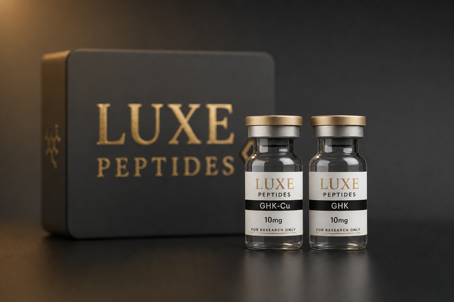
For laboratory research use only. GHK-Cu is not a drug, dietary supplement, or cosmetic ingredient supplied for human or veterinary use, and nothing below is a recommendation for administration to humans or animals.
GHK-Cu is the copper(II) coordination complex of the tripeptide glycyl-L-histidyl-L-lysine (GHK). The peptide sequence was first isolated from human plasma albumin fractions in the early 1970s, and the copper complex has since become one of the most extensively characterized small peptide–metal systems in extracellular matrix and dermal fibroblast research. This overview summarizes what preclinical literature reports about its chemistry and studied mechanisms, and how the copper-bound and copper-free forms differ as laboratory materials.
Structure and copper coordination
GHK is a three-residue peptide (Gly-His-Lys) with a molecular weight of roughly 340 Da. Its architecture gives it an unusually well-defined metal-binding site: the N-terminal amine, the imidazole nitrogen of histidine, and a deprotonated backbone amide nitrogen together form a square-planar chelation geometry around Cu(II), with the lysine side chain remaining free and positively charged at physiological pH. The resulting 1:1 complex has a molecular weight near 402 Da and is visibly blue-violet in aqueous solution.
Because the affinity of GHK for Cu(II) is high but not irreversible, the literature discusses the complex primarily as a copper shuttle rather than a copper sink — a carrier able to exchange copper with plasma proteins and with cellular uptake machinery. That framing matters mechanistically, because several enzymes involved in matrix assembly and redox regulation are cuproenzymes, including lysyl oxidase and Cu/Zn superoxide dismutase. Researchers investigating GHK-Cu therefore generally treat copper delivery and peptide signaling as two coupled variables rather than one, and copper-free GHK is frequently run as a comparator for exactly this reason.
Reported plasma concentrations of the free tripeptide are often cited as declining with donor age — on the order of 200 ng/mL in young adults versus roughly 80 ng/mL by the sixth decade. This observation is descriptive rather than causal, but it is the origin of much of the interest in the sequence as an endogenous signaling fragment.
Mechanisms studied in preclinical research
Extracellular matrix turnover
The earliest mechanistic work examined cultured fibroblasts. Maquart and colleagues reported in the late 1980s that GHK-Cu increased collagen synthesis in fibroblast culture at nanomolar to micromolar concentrations. Later work from the same group examined proteoglycan and glycosaminoglycan expression, reporting changes in decorin and related small proteoglycans in wound-model tissue exposed to the complex. Decorin is of mechanistic interest because it participates in collagen fibril organization and in sequestering TGF-β, placing it upstream of matrix architecture rather than simply being a structural output.
A related line of research examined matrix metalloproteinases. Siméon and colleagues reported that GHK-Cu modulated MMP-2 alongside its tissue inhibitors TIMP-1 and TIMP-2 in fibroblast culture. Because both the protease and its inhibitors moved, the reported profile is generally interpreted as increased matrix turnover — coordinated breakdown and resynthesis — rather than one-directional accumulation or degradation. This is the mechanistic claim most consistently supported across the in vitro literature.
Gene-expression profiling
Interest broadened considerably after transcriptomic datasets became available. Pickart and colleagues analyzed GHK against reference expression signatures using the Broad Institute Connectivity Map and reported that the peptide was associated with modulation of several thousand human genes in cultured cells at low-micromolar exposures, with enrichment in categories related to matrix remodeling, DNA repair, ubiquitin–proteasome activity, and inflammatory signaling. These are correlative signature analyses, not demonstrations of a single receptor or pathway, and no canonical GHK receptor has been established. Researchers designing follow-up work generally treat the gene-signature literature as hypothesis-generating.
Redox and inflammatory markers
GHK-Cu has been described in vitro as possessing superoxide-dismutase-like activity, consistent with the redox behavior of a chelated Cu(II) center. Separate cell-culture reports have examined effects on inflammatory cytokine markers such as TNF-α and IL-6 in fibroblast systems. Copper chemistry cuts both ways here: chelated copper can attenuate free-radical chemistry at low concentrations while free or loosely bound copper can catalyze Fenton-type reactions. Concentration and complex integrity are therefore critical experimental variables, and studies that do not report copper speciation are difficult to interpret.
Animal wound-model work
Topical GHK-Cu has been evaluated in rodent, rabbit, and canine wound models, where investigators have reported differences in closure kinetics and granulation-tissue characteristics relative to vehicle controls. These studies are small, heterogeneous in formulation and endpoint definition, and not consistently blinded, so they are best read as supporting the matrix-turnover mechanism rather than as outcome evidence.
GHK versus GHK-Cu as research materials
Both forms are sold and studied, and they are not interchangeable in an assay. The table below summarizes the practical differences.
| Property | GHK (copper-free) | GHK-Cu (copper complex) |
|---|---|---|
| Composition | Gly-His-Lys tripeptide, typically as acetate or TFA salt | Gly-His-Lys coordinated 1:1 to Cu(II) |
| Approx. molecular weight | ~340 Da (free base) | ~402 Da (1:1 complex) |
| Appearance in solution | Colorless | Blue to blue-violet |
| Copper contribution | None; depends on copper already present in the medium | Delivers copper stoichiometrically with the peptide |
| Typical research role | Isolating peptide-attributable signaling from metal effects | Studying combined peptide + copper-delivery mechanisms |
| Handling sensitivity | Standard peptide handling | Additionally sensitive to pH extremes, chelators, and light |
Product pages for both forms are here: GHK-Cu and GHK (basic).
Laboratory handling and solution preparation
Lyophilized material is normally stored cold, desiccated, and protected from light, with reconstituted stock treated as short-lived. For the copper complex, color is a useful qualitative indicator: loss of the characteristic blue tint, or precipitate formation, suggests copper dissociation or degradation of the complex and argues for preparing fresh stock rather than proceeding. Strongly alkaline buffers and competing chelators such as EDTA will perturb the complex and should be accounted for in buffer selection.
Concentration work is ordinary laboratory arithmetic: diluent volume divided into peptide mass gives the stock concentration, from which working dilutions follow. The reconstitution calculator handles this conversion. Note that for GHK-Cu, molar calculations should use the complex mass, not the free-peptide mass — a roughly 18% difference that is easy to overlook and that shifts every downstream molarity.
Limitations of the evidence base
Three caveats recur across this literature. First, the great majority of mechanistic data comes from fibroblast culture and small-animal models; extrapolation beyond those systems is not supported. Second, no defined receptor has been identified, so “mechanism” here means observed pathway and expression changes, not a resolved signaling cascade. Third, results are sensitive to copper speciation, peptide purity, and salt form — which is why analytical documentation matters. Batch-level identity and purity data are published in the COA library.
Reviewed for research accuracy: July 30, 2026.
References
- Pickart L, Thaler MM. Nature New Biology. 1973;243:85–87. (Original isolation of the GHK sequence from human plasma.)
- Maquart FX, Pickart L, Laurent M, Gillery P, Monboisse JC, Borel JP. FEBS Letters. 1988;238(2):343–346.
- Siméon A, Emonard H, Hornebeck W, Maquart FX. Life Sciences. 2000;67(18):2257–2265.
- Siméon A, Monier F, Emonard H, et al. Journal of Investigative Dermatology. 2000. (Glycosaminoglycan and small-proteoglycan expression in wound models exposed to the tripeptide–copper complex.)
- Pickart L, Vasquez-Soltero JM, Margolina A. BioMed Research International. 2015;2015:648108. DOI: 10.1155/2015/648108.
- Pickart L, Margolina A. International Journal of Molecular Sciences. 2018;19(7):1987. PMID: 29986520. DOI: 10.3390/ijms19071987.
- Cangul IT, Gul NY, Topal A, Yilmaz R. BMC Veterinary Research. 2006;2:29. (Comparative open-wound model evaluation of a tripeptide–copper complex.)
- Additional preclinical reports have examined GHK and GHK-Cu effects on inflammatory cytokine markers in fibroblast culture; these are small single-laboratory studies and are cited here generically.
Research use only. All compounds discussed are supplied strictly for in vitro laboratory and preclinical research by qualified personnel, and are not for human consumption, therapeutic use, cosmetic use, or veterinary application.